Black stain is characterized by a dark line or an incomplete coalescence of dark dots localized on the cervical third of the tooth. Over the past century, its etiology has been widely debated. Based on studies published between 2001 and 2014, the prevalence of black stain ranges from 2.4% to 18%, with no significant difference between sexes.
Most studies indicate a correlation between the presence of black stain and a lower incidence of dental caries. The microflora associated with black stain is predominantly composed of Actinomyces species and demonstrates lower cariogenic potential compared to nondiscolored dental plaque. The dark coloration is believed to result from iron or copper and sulfur complexes. Additionally, individuals with black stain tend to have higher salivary calcium levels and greater buffering capacity. Contributing factors may include dietary habits, socioeconomic status, and iron supplementation.
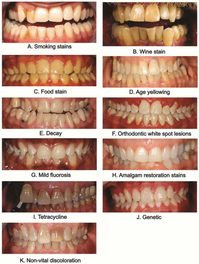
1. Introduction
Tooth discoloration can be classified based on its location into:
- Extrinsic discoloration: Deposited on the tooth surface or acquired pellicle
- Intrinsic discoloration: Occurs within the tooth structure, typically during development
- Internalized discoloration: Results from incorporation of external stains into the tooth after development
Black stain is a specific type of extrinsic discoloration. It appears as a dark line or series of dots along the cervical third of the tooth, following the contour of the gingival margin. It is firmly attached to the tooth surface.
Although more commonly observed in children, black stain can also occur in adults, with studies indicating equal prevalence among males and females. It is considered a form of dental plaque with a tendency toward calcification.
2. Nature and Microbiology of Black Stain
Black stain consists of microorganisms embedded within a matrix. Ultrastructural studies reveal that most bacteria present are Gram-positive rods, predominantly Actinomyces species.
Recent molecular analyses have shown:
- Higher levels of Actinomyces naeslundii
- Lower levels of Lactobacillus species compared to nondiscolored plaque
The pigment responsible for the black coloration is believed to be ferric sulfide, formed by the interaction between hydrogen sulfide (produced by bacteria) and iron in saliva or gingival fluid.
Chemical analysis has also demonstrated:
- Higher calcium and phosphate content
- Presence of sulfur and iron/copper complexes contributing to pigmentation
3. Black Stain Prevalence and Caries Status
The prevalence of black stain varies between 2.4% and 18%, largely due to differences in diagnostic criteria and study populations.
Key Findings from Epidemiological Studies
- Several studies indicate that children with black stain exhibit lower caries prevalence
- Some studies report no significant association between black stain and caries
- In both children and adults, reduced DMFT/dmft scores have been observed in individuals with black stain
These findings suggest that the reduced caries risk is likely due to overall lower caries activity rather than localized protection at stained sites.
4. Factors Contributing to the Formation of Black Stain
The exact etiology of black stain remains unclear. However, several contributing factors have been proposed:
4.1 Demographic Factors
- No consistent association with sex
- Possible increase in prevalence with age
4.2 Dietary Habits
- Increased consumption of:
- Vegetables
- Fruits
- Dairy products
- Eggs
- Soy-based products
4.3 Feeding and Water Source
- Higher occurrence in children not bottle-fed
- Possible association with consumption of tap water
4.4 Oral Hygiene Practices
- Conflicting evidence regarding the role of oral hygiene
- Some studies suggest fluoride products may promote staining
4.5 Socioeconomic Status
- Mixed findings regarding parental education and socioeconomic background
4.6 Iron Supplementation
- Iron intake during pregnancy or childhood may contribute to stain formation
5. Chemical Composition of Black Stain
Biochemical studies have identified the following characteristics:
- Higher levels of calcium and phosphate
- Presence of ferric sulfide as the primary pigment
- Detection of iron, copper, and sulfur complexes
Advanced analytical methods suggest that metallic ions and sulfur compounds interact to produce the characteristic dark color. It is also noted that sample collection methods may influence the detection of metallic elements.
6. Salivary Parameters
Saliva plays a critical role in maintaining oral health and preventing dental caries.
In individuals with black stain, studies have reported:
- Higher salivary pH
- Increased buffering capacity
- Elevated calcium and phosphate levels
- Higher total protein concentration
- Lower glucose levels
These factors contribute to a less cariogenic oral environment.
7. Microbiology of Black Stain
Black stain is considered a specialized form of dental plaque with distinct microbial composition.
Key Microbiological Features
- Predominance of Gram-positive rods
- High prevalence of Actinomyces species
- Lower levels of cariogenic bacteria such as Streptococcus mutans and Lactobacillus
Some studies also report:
- Absence of certain periodontal pathogens
- Variability in bacterial composition across populations
Overall, the microbial profile of black stain is associated with reduced cariogenic potential.
8. Conclusion
Black stain is a form of extrinsic tooth discoloration typically appearing along the gingival margin. Its prevalence ranges from 2.4% to 18%, with no significant gender predilection.
Key characteristics include:
- Dominance of Actinomyces species
- Lower presence of cariogenic bacteria
- Association with higher salivary calcium and buffering capacity
- Possible protective relationship with dental caries
Although the exact etiology remains unclear, current evidence suggests that black stain may be linked to a less cariogenic oral environment. Further research is required to better understand its formation and potential protective role against dental caries.
Conflict of Interest
The authors declare no conflict of interest regarding the publication of this article.
References
- Hattab FN, Qudeimat MA, Al-Rimawi HS. Dental discoloration: an overview. Journal of Esthetic Dentistry. 1999;11(6):291–310.
DOI: https://doi.org/10.1111/j.1708-8240.1999.tb00413.x - Watts A, Addy M. Tooth discolouration and staining: a review of the literature. British Dental Journal. 2001;190(6):309–316.
DOI: https://doi.org/10.1038/sj.bdj.4800959 - Reid JS, Beeley JA, MacDonald DG. Investigations into black extrinsic tooth stain. Journal of Dental Research. 1977;56(8):895–899.
DOI: https://doi.org/10.1177/00220345770560081001 - Chen X, Zhan JY, Lu HX, et al. Factors associated with black tooth stain in Chinese preschool children. Clinical Oral Investigations. 2014;18(9):2059–2066.
DOI: https://doi.org/10.1007/s00784-013-1184-z - Garcia Martin JM, Garcia MG, Leston JS, Pendas SL, Martin JJ, Garcia-Pola MJ. Prevalence of black stain and associated risk factors in preschool Spanish children. Pediatrics International. 2013;55(3):355–359.
DOI: https://doi.org/10.1111/ped.12066 - Reid JS, Beeley JA. Biochemical studies on the composition of gingival debris from children with black extrinsic tooth stain. Caries Research. 1976;10(5):363–369.
DOI: https://doi.org/10.1159/000260217 - Theilade J, Slots J, Fejerskov O. The ultrastructure of black stain on human primary teeth. Scandinavian Journal of Dental Research. 1973;81(7):528–532.
DOI: https://doi.org/10.1111/j.1600-0722.1973.tb00360.x - Slots J. The microflora of black stain on human primary teeth. Scandinavian Journal of Dental Research. 1974;82(7):484–490.
DOI: https://doi.org/10.1111/j.1600-0722.1974.tb00405.x - Heinrich-Weltzien R, Bartsch B, Eick S. Dental caries and microbiota in children with black stain and non-discoloured dental plaque. Caries Research. 2014;48(2):118–125.
DOI: https://doi.org/10.1159/000353469 - Tantbirojn D, Douglas WH, Ko CC, McSwiggen PL. Spatial chemical analysis of dental stain using wavelength dispersive spectrometry. European Journal of Oral Sciences. 1998;106(5):971–976.
DOI: https://doi.org/10.1046/j.0909-8836..t01-8-.x - Surdacka A. Amount and pH of the saliva in children and adolescents with black tartar. Czasopismo Stomatologiczne. 1989;42(6):381–386.
- Surdacka A. Chemical composition of the saliva in children and adolescents with black tartar. Czasopismo Stomatologiczne. 1989;42(10–12):525–533.
- Aysun G, Akyüz S, Öztürk LK, Yarat A. Salivary parameters and caries indices in children with black tooth stains. Journal of Clinical Pediatric Dentistry. 2012;36(3):285–288.
DOI: https://doi.org/10.17796/jcpd.36.3.21466m672t723713 - Heinrich-Weltzien R, Monse B, van Palenstein Helderman W. Black stain and dental caries in Filipino schoolchildren. Community Dentistry and Oral Epidemiology. 2009;37(2):182–187.
DOI: https://doi.org/10.1111/j.1600-0528.2008.00458.x - Koch MJ, Bove M, Schroff J, Perlea P, García-Godoy F, Staehle HJ. Black stain and dental caries in schoolchildren in Potenza, Italy. Journal of Dentistry for Children. 2001;68(5–6):353–355.
- Boka V, Trikaliotis A, Kotsanos N, Karagiannis V. Dental caries and oral health-related factors in a sample of Greek preschool children. European Archives of Paediatric Dentistry. 2013;14(6):363–368.
DOI: https://doi.org/10.1007/s40368-013-0097-5 - França-Pinto CC, Cenci MS, Correa MB, et al. Association between black stains and dental caries in primary teeth: findings from a Brazilian population-based birth cohort. Caries Research. 2012;46(2):170–176.
DOI: https://doi.org/10.1159/000337280 - Gasparetto A, Conrado CA, Maciel SM, Miyamoto EY, Chicarelli M, Zanata RL. Prevalence of black tooth stains and dental caries in Brazilian schoolchildren. Brazilian Dental Journal. 2003;14(3):157–161.
DOI: https://doi.org/10.1590/s0103-64402003000300003 - Bhat S. Black tooth stain and dental caries among Udaipur school children. International Journal of Public Health Dentistry. 2010;1:13–15.
- Pickerill HP. A sign of immunity. British Dental Journal. 1923;2:967–968.
- Panagidis D, Schulte AG. Caries prevalence in 12-year-old Cypriot children. Community Dental Health. 2012;29(4):297–301.
DOI: https://doi.org/10.1922/CDH_2774Panagidis05 - Shmuly T, Zini A, Yitschaky M, Yitschaky O. Can black extrinsic tooth discoloration predict a lower caries score rate in young adults? Quintessence International. 2014;45(5):439–444.
DOI: https://doi.org/10.3290/j.qi.a31535
